Patient
Height (cm)
Sex
- select -
Male
Female
Reset
Agents
Drug
Conc.
Vol (mL)
Dose given
Safe vol. remaining
Add drug
- select -
Lignocaine
Lignocaine + Adr
Bupivacaine
Ropivacaine
Concentration
-
Add row
Cumulative Toxic Fraction
Used: 0.0%
Remaining: 100.0%
CTF: each drug's entered volume is expressed as a fraction of its absolute maximum dose at the selected weight. Fractions sum across all agents. Safe vol. remaining shows the maximum additional volume of each drug before CTF reaches 1.0. Always verify independently - not a substitute for clinical judgement.
Enter patient age or weight to view doses
00:00:00
Session
00:00
Start
Reset
Patient
Total body weight
—
kg (entered)
Ideal body weight
—
Devine formula
Lean body weight
—
Janmahasatian
Disclaimer & Licence
Clinical use. Hypnos Vantage is a reference aid only. All drug doses, calculations, and clinical information must be independently verified against current local guidelines, formulary, and product information before use in patient care. The author accepts no responsibility for clinical decisions made in reliance on this tool.
Licence. © 2025 Hypnos Medical. Licensed under
CC BY-NC 4.0
— free to use, share, and adapt with attribution. Commercial use is prohibited without explicit permission.
Abdomen
Gastric US
Lung
TTE
Venous
View Summary
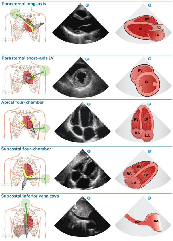
View Window Structures seen Key uses PLAX Parasternal, 3rd–4th ICS LSE RV, LV, AO, LA, MV, AV LV size/function, valves, pericardium PSAX LV Parasternal, rotate 90° RV, LV (short axis “D-sign”) LV wall motion, RV pressure/volume overload A4C Apical, cardiac apex lateral chest RV, LV, RA, LA, MV, TV Biventricular function, valves, filling SC4C Subcostal, below xiphoid RV, LV, RA, LA, pericardium Tamponade, RV function, pericardial effusion SC IVC Subcostal, rotate to midline IVC, RA junction Volume status, right heart pressure
Probe Selection & Patient Positioning
Probe selection
Linear (high-frequency): best for pleural line, pneumothorax, B-line counting, diaphragm thickness
Curvilinear (low-frequency): pleural effusion, consolidation, diaphragm excursion, deep artefacts
Phased array (small footprint): useful in narrow anterior intercostal spaces
Patient positioning
Supine: anterior zones — probe anteriorly for pneumothorax (air rises)
Semi-recumbent: apex view; clavicles can limit access
Posterolateral: dependent pathology — consolidation, effusion, haemothorax
6-zone protocol
Scan bilaterally across 6 zones, 3 intercostal spaces per zone:
Anterior zones: 2nd–3rd ICS mid-clavicular line (upper & lower BLUE points)
Lateral zones: anterior–posterior axillary line, at the level of the axilla
Posterior zones: posterior axillary line, above and below the spine of scapula
Artefact Terminology
Term Description Significance A-lines Horizontal reverberation artefacts below pleural line at regular intervals (= probe-to-pleura distance) Normal aerated lung or pneumothorax B-lines ≥3 vertical laser-like artefacts from pleural line to far field, erasing A-lines Interstitial fluid/oedema • ≥3 per zone = pathological C-lines Hypoechoic subpleural comet-tail artefacts Consolidation E-lines Vertical hyperechoic lines from thoracic wall (not pleura) Subcutaneous emphysema Lung sliding Visceral/parietal pleura sliding (“marching ants” on B-mode) Absent in pneumothorax, adhesions, post-pleurodesis Seashore sign M-mode: granular “sand” below static “sea” of chest wall Normal lung sliding present Barcode sign M-mode: uniform horizontal lines (no sliding) Pneumothorax Lung point Transition between absent and present sliding 100% specific for pneumothorax Lung pulse Cardiac pulsation transmitted to pleural line Lung not ventilated (endobronchial intubation) • absent in pneumothorax Spine sign Thoracic vertebrae visible on US Pleural effusion or haemothorax present (normally hidden by aerated lung) Curtain sign Cranio-caudal movement of lung base during respiration Reduced/absent: effusion, atelectasis, or basal consolidation Air bronchogram Aerated bronchi in airless lung Dynamic = pneumonia • Static = resorptive atelectasis Lung hepatisation Lung tissue isoechoic to liver parenchyma Substantial consolidation Lung monster Atelectatic lung as wedge-shaped mass moving with respiration within effusion Large pleural effusion
Ref: Algain et al., ATOTW 523, WFSA 2024
Pathology Profiles
Pneumothorax
Absent lung sliding in B-mode • Barcode sign replaces seashore sign in M-mode
Lung point (100% specific) — seek actively if sliding absent
Also absent in: endobronchial intubation, pleuro-parenchymal adhesions, subpleural bullae, post-pleurodesis
Lung sliding sensitive but NOT specific for pneumothorax
Pleural Effusion
Anechoic space between pleural layers at posterolateral scan • Detects as little as 5–20 mL
Spine sign positive (vertebrae visible through fluid)
Large effusion: lung monster sign (atelectatic lung moves with respiration within fluid)
Use acoustic window of liver (right) or spleen (left) at costo-phrenic angle
Pulmonary Oedema
≥3 B-lines per zone (convex probe) or ≥6 B-lines (linear probe) = pathognomonic
Multiple bilateral B-lines; may precede radiographic changes
Number of B-lines correlates with severity; resolution correlates with clinical improvement
Consolidation / Pneumonia
Hepatisation: hyperechoic heterogeneous region resembling liver parenchyma
Air bronchogram: branching tubular structures within consolidated tissue
Dynamic bronchogram (propagates with breathing): specificity 94%, PPV 97% for pneumonia
Static bronchogram: isolated trapped air → resorptive atelectasis
Vascular flow on colour Doppler within consolidation confirms pneumonia
ARDS
Multiple B-lines with irregular pleural line pattern
Small posterior and basal consolidations • Air bronchogram
Bilateral non-homogeneous distribution distinguishes from cardiogenic oedema
Subcutaneous Emphysema
E-lines: multiple vertical hyperechoic lines from thoracic wall to pleural line
E-lines obscure the pleural line; bat sign may be lost
Associated with pneumothorax — scan other regions for lung point
BLUE Protocol — Acute Respiratory Failure
3-minute protocol. Diagnoses cause of acute respiratory failure with 90.5% accuracy .
Landmark identification
Upper BLUE point: base of 2nd and 3rd fingers — place 4 fingers (excluding thumb) below clavicle with fingertips on sternum
Lower BLUE point: centre of palm of right hand placed below left with forefingers touching
Mirror for left side • Scan lung bases only if upper/lower BLUE points non-diagnostic
PLAPS point: from lower BLUE point, move to posterior axillary line at thoracoabdominal border
Profile Findings Likely Diagnosis A-profile Bilateral A-lines + lung sliding Asthma or COPD exacerbation (if no DVT) • PE (if DVT) B-profile Bilateral anterior B-lines + lung sliding Pulmonary oedema B’-profile Bilateral B-lines + absent lung sliding Pneumonia A/B-profile Asymmetric: A-lines one side, B-lines other Pneumonia C-profile Anterior consolidation Pneumonia A-profile + PLAPS A-lines anteriorly + posterior consolidation/effusion Pneumonia A-profile + no sliding Absent sliding + barcode sign ± lung point Pneumothorax
Ref: Lichtenstein & Mazière, Chest 2008. Algain et al., ATOTW 523, WFSA 2024.
Diaphragm Assessment
Technique
Excursion (M-mode, curvilinear): subxiphoid view, measure caudal movement during deep inspiration
Thickness (linear): 9th ICS anterior axillary line • measure end-inspiration vs end-expiration
Thickening fraction (TFdi ) = (thicknessinsp − thicknessexp ) / thicknessexp
Measurement Normal (male) Normal (female) Abnormal Quiet breathing excursion 1.8 ± 0.3 cm 1.6 ± 0.3 cm Deep breathing excursion 7.0 ± 0.6 cm 5.7 ± 1.0 cm Sniff test excursion 2.9 ± 0.6 cm 2.6 ± 0.5 cm Thickness at end-expiration >0.2 cm >0.2 cm <0.2 cm = atrophy TFdi ≥20% ≥20% <20% = paresis/paralysis
Phrenic nerve palsy
Partial palsy: sniff test shows 25–75% reduction in caudal movement
Complete palsy: paradoxical cephalad movement or ≥75% reduction
High sensitivity (93%) and specificity (100%) for phrenic nerve dysfunction
eFAST — Overview
What is eFAST?
Extended Focused Assessment with Sonography in Trauma — adds bilateral lung views to the original FAST exam
Detects: haemoperitoneum, haemopericardium, haemothorax, pneumothorax
Goal: identify life-threatening free fluid in <5 minutes
Positive eFAST in an unstable patient → immediate surgical intervention
Probe & setup
Curvilinear (3–5 MHz) for all abdominal views • Low-frequency phased array as alternative
Linear (high-frequency) for lung views (pneumothorax)
Patient supine throughout • Scan from patient’s right side
Sagittal views: indicator to head • Transverse views: indicator to patient’s right
Full bladder improves pelvic views — scan before catheter placement if possible
Trendelenburg (5°) improves RUQ/LUQ sensitivity; Reverse Trendelenburg improves pelvic views
eFAST Views
View Probe position Structures Free fluid location RUQ Posterior–mid axillary line, 8th–11th ICS, indicator cephalad Liver, right kidney, diaphragm, R hemithorax Morrison's pouch (hepatorenal space) • Liver tip/paracolic gutter • Subphrenic space • R pleural spaceLUQ Posterior axillary line, 6th–9th ICS, more cephalad & posterior than RUQ Spleen, left kidney, diaphragm, L hemithorax Perisplenic space • Splenorenal recess • Subphrenic space • L pleural space Pelvis (long) Midline just above pubic symphysis, indicator cephalad, angled caudally Bladder (long axis), uterus/prostate Rectovesical pouch (M) • Pouch of Douglas (F) • Vesicouterine pouch (F) Pelvis (transverse) Midline above pubis, rotate 90°, indicator to patient’s right Bladder, uterus/prostate, rectum Lateral to bladder • Posterior to uterus in females Cardiac (SC) Subxiphoid, indicator to patient’s right, beam towards left shoulder 4 chambers, pericardium Pericardial effusion/tamponade — anechoic layer around heart Lungs (bilateral) Anterior chest, 2nd–3rd ICS mid-clavicular (linear probe) Pleural line, A-lines, lung sliding Absent sliding → pneumothorax • Barcode sign on M-mode
RUQ most sensitive overall; fluid around liver tip has highest sensitivity (>93%). LUQ technically most challenging.
Interpreting Free Fluid
Positive exam — any of:
Anechoic (black) stripe in any dependent peritoneal space
Morrison's pouch: stripe between liver and kidney — even 1–2 mm is significant
Perisplenic: fluid around spleen (most common LUQ location — NOT splenorenal, due to splenorenal ligament)
Pelvic: fluid posterior to bladder (males) or posterior to uterus — Pouch of Douglas (females)
Above diaphragm: anechoic area cephalad to liver or spleen = haemothorax
Pericardial: anechoic layer surrounding heart = tamponade until proven otherwise
Pitfalls & mimics
Gallbladder, hepatic veins, biliary ducts → can mimic RUQ free fluid
Edge artefact → false positive at Morrison’s pouch interface
Renal cysts, perinephric fat → can resemble free fluid
Seminal vesicles (males), ovarian cysts, physiologic pelvic fluid (females) → false positive pelvis
Pre-existing ascites or peritoneal dialysis fluid → may be indistinguishable from haemorrhage
Ultrasound cannot differentiate blood from ascites or urine — always correlate clinically
eFAST cannot localise the source of bleeding — CT required if patient stable
Performance characteristics
Detection threshold: ~200 mL in most spaces; pelvic and costophrenic angles most sensitive for small volumes
Overall sensitivity ~69–85%; NPV 97% with serial exams
Serial exams reduce false negative rate by up to 50%
Haemothorax sensitivity 92–100%, specificity 92–100%
~30 supervised exams recommended to achieve competency
Beyond FAST — Other Abdominal Applications
Abdominal aorta
Indication: suspected AAA in haemodynamically unstable patient or back/abdominal pain
Transverse and longitudinal views from xiphoid to umbilicus
Normal aortic diameter <3 cm • ≥3 cm = aneurysm • ≥5.5 cm or rapid expansion = surgical threshold
Free fluid around aorta = rupture until proven otherwise
Renal (hydronephrosis)
Indication: sepsis with possible obstructive cause, oliguria/anuria, flank pain
Probe posterolateral, long axis of kidney • Look for dilated calyces and renal pelvis
Hydronephrosis: anechoic branching structure (fluid) within echogenic renal sinus
Bladder volume assessment: transverse & longitudinal views above pubic symphysis
Biliary
Indication: RUQ pain, sepsis of biliary origin
Subcostal probe, long axis of gallbladder • Assess wall thickness, stones, pericholecystic fluid, CBD diameter
Cholecystitis: wall >3 mm, pericholecystic fluid, gallstones, sonographic Murphy’s sign
CBD dilation: >6 mm (or >8 mm post-cholecystectomy) suggests biliary obstruction
Refs: StatPearls (FAST); Arora et al. in POCUS in Critical Care, Anesthesia and Emergency Medicine (Springer, 2024); Dhir et al. Cureus 2024.
Venous POCUS — content coming soon
Indications
Use gastric US when
Prandial status uncertain — cognitive dysfunction, language barrier, unclear history, paediatric
Delayed gastric emptying suspected — diabetes, CKD, acute pain, opioids, obesity, pregnancy, GLP-1 agonist use (within 4 weeks)
Key threshold
Empty antrum or <1.5 mL/kg clear fluid → consistent with fasting state (low aspiration risk)
≥1.5 mL/kg clear fluid OR any solids → consistent with full stomach (high aspiration risk)
Image Acquisition
Transducer: Low-frequency curved array (1–5 MHz). Paeds/low BMI: high-frequency linear (5–12 MHz).
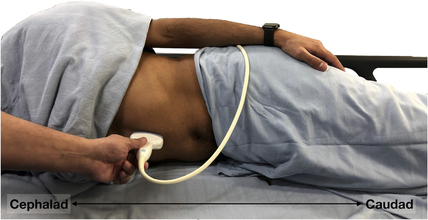
Position: Right lateral decubitus — most sensitive. Supine also used. Semi-recumbent if right lateral decubitus not possible.
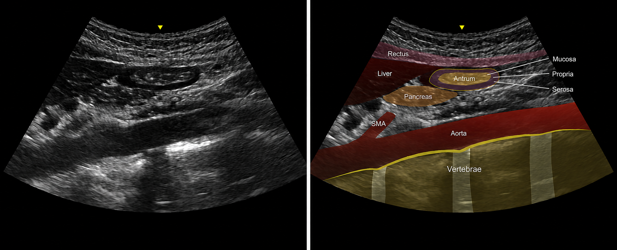
Technique: Sagittal plane, epigastrium just below xiphisternum. Sweep left to right seeking: vertebral bodies → aorta → SMA → pancreas → left lobe liver → gastric antrum in short axis. Heel-to-toe to minimise obliquity.
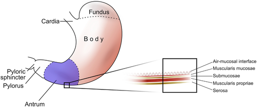
Fig 1. Gastric sections and antral wall layers
Fig 2. Right lateral decubitus position, transducer in epigastrium
El-Boghdadly et al., BJA Education 2019
Image Interpretation
Empty stomach — LOW RISK
Antrum small, flat, collapsed in both supine and right lateral decubitus
‘Bull’s-eye’ appearance when round/ovoid
Walls appear thick; muscularis mucosae prominent
Diagnosis of empty antrum requires right lateral decubitus position after continued observation
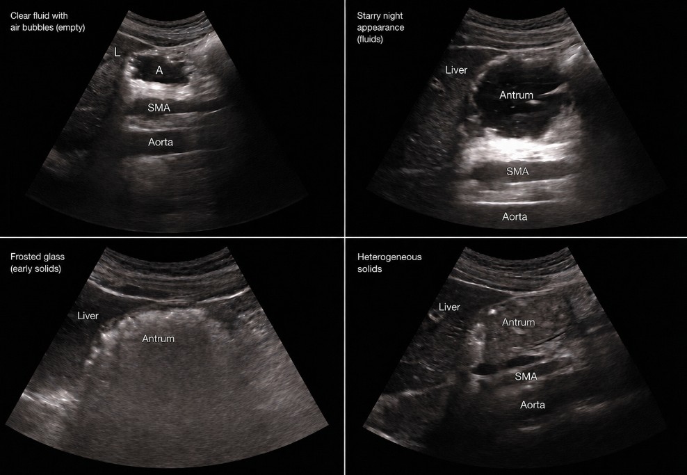
Clear fluid — interpret with volume
Antrum distended, thin-walled, hypoechoic content
Air bubbles may appear as hyperechoic dots (‘starry night’ appearance)
Grade 0: empty in both supine and right lateral decubitus → low risk (45–50% of fasted patients)
Grade 1: empty supine, fluid in right lateral decubitus → <1.5 mL/kg → low risk (45–50%)
Grade 2: fluid in both supine and right lateral decubitus → ≥1.5 mL/kg → HIGH RISK (3–5%)
Solids — HIGH RISK
Early: ‘frosted glass’ appearance — air from chewing obscures deep structures
Later: hyperechoic, heterogeneous content; distended antrum; peristalsis visible
Thick fluids (milk, yoghurt): homogeneous hyperechoic; possible biphasic if curdled
ANY solid content = high risk regardless of quantity
El-Boghdadly et al., BJA Education 2019
ALS
Acute Stroke
Anaphylaxis
Aneurysm Rupture (IR)
Aneurysm Rupture (OR)
Autonomic Hyperreflexia
Bradycardia
Bronchospasm
Failure to Wake
High Airway Pressure
Hypertension
Hypotension
Increased ICP
LA Toxicity
Loss of Evoked Potentials
Malignant Hyperthermia
Seizures
Tachycardia
Vasospasm
Venous Air Embolism
Adult Advanced Life Support (ANZCOR 2021)
Based on ANZCOR guidelines. Always follow current local protocols.
UNRESPONSIVE + NOT BREATHING NORMALLY
Call for help • Start CPR • Attach defibrillator
CPR Quality
Rate: 100-120/min 5-6 cm 30:2 (or continuous if intubated)
Shockable (VF/pVT)
Shock: 200 J biphasic 3-5 min
Non-Shockable (PEA/Asystole)
Adrenaline 1mg IV as soon as access3-5 min
Reversible Causes (4H + 4T)
Hypoxia • Hypovolaemia
Post-ROSC Care
SpO2 94-98% • EtCO2 35-45 mmHg • MAP ≥65 mmHg • 12-lead ECG (cath lab if STEMI)
Ref: ANZCOR Guideline 11 (2021) • Resuscitation Council UK 2021
Local Anaesthetic Systemic Toxicity (LAST)
AAGBI/ANZCA guidelines. Lipid Emulsion Therapy is the definitive treatment.
Recognition — Signs & Symptoms
CNS (early): Perioral tingling, metallic taste, tinnitus, agitation, confusion, drowsiness
CVS: Hypotension, bradycardia, VT/VF, asystole
CNS (severe): Loss of consciousness, seizures, respiratory arrest
Note: CVS collapse may occur without preceding CNS signs (esp. bupivacaine)
Immediate Actions
1. Stop injecting LA immediately Start Intralipid 20% immediately
💉 Intralipid 20% (Lipid Emulsion Therapy)
Bolus
1.5 ml/kg IV over 1 min(70 kg adult = ~100 ml ) 2 more times at 5 min intervals if no response
Infusion
15 ml/kg/hr (70 kg adult = ~1000 ml/hr ) 12 ml/kg
Location of Intralipid at WRH: Confirm with your department
If Cardiac Arrest
Standard ALS • Start Intralipid immediately • Avoid vasopressin≤1 mcg/kg (small doses)
Ref: AAGBI LAST guidelines 2023 •
lipidrescue.org • ANZCA crisis management cards
Malignant Hyperthermia (MH)
MH Hotline (MHAUS): +1 800 986 4287 • MH NZ contact: confirm with your department
Recognition
Early: Unexplained rise in EtCO2, tachycardia, masseter spasm (after sux), tachypnoeaTriggers: Volatile anaesthetic agents, succinylcholine
Late: Rising temp (>38.5°C and rising), rigidity, dark urine (myoglobinuria), metabolic acidosis, hyperkalaemia
Immediate Actions
1. Stop all trigger agents immediately IV agents only (propofol, opioids, NMBA)
💉 Dantrolene (definitive treatment)
Initial Dose
2.5 mg/kg IV rapid bolus(70 kg adult = ~175 mg = 9 vials )
Repeat Dosing
Repeat 1 mg/kg every 5-10 min10 mg/kg 24-48h
Supportive Treatment
Cooling: Ice packs (axilla, groin, neck), cold IV fluids, bladder irrigation, surface cooling<38.5°C • Stop active cooling at 38°C
Hyperkalaemia: Calcium chloride, sodium bicarbonate, dextrose/insulinArrhythmia: Amiodarone (avoid CCBs — interact with dantrolene)Renal: Forced diuresis (urine output >1 ml/kg/hr) for myoglobinuria
Monitoring & Post-Acute Care
ABG, electrolytes, CK, LFTs, coagulation • Urine myoglobin • Temperature (core & peripheral)
Ref: ANZCA/MHAUS MH guidelines •
mhaus.org • European MH Group guidelines 2020
Tachycardia
NZ Anaesthetic Crisis Handbook (Hollingworth) & ANZCOR 2021.
Immediate Assessment
Check: Is the patient oxygenated & anaesthetised? (A, B, C, D, E)
If MAP <65 + HR >150 with diagnostic uncertainty → synchronised DC cardioversion immediately
Differentiate: sinus tachycardia vs complex tachy-arrhythmia
ECG: Is rhythm regular? QRS narrow or wide?
Sinus Tachycardia — Treat the Cause
Hypovolaemia • Pain / light anaesthesia • Drug error • Sepsis • MH • Thyroid storm • Electrolyte abnormality • Tamponade • Tension PTX
Complex Tachy-arrhythmia (MAP >65 mmHg) — by rhythm
Regular narrow (SVT): Vagal manoeuvres → Adenosine 6 mg rapid IV bolus + saline flush; no response: 12 mg; repeat 12 mg; or metoprolol 2.5 mg boluses (max 15 mg)
Regular wide (VT): Amiodarone 300 mg slow IV push • If SVT with aberrancy: treat as narrow
Irregular narrow (AF): Metoprolol 2.5 mg boluses titrated; or amiodarone 300 mg slow push
Irregular wide — torsades: Magnesium 10 mmol (5 ml of 49.3%) IV over 2 min
AF + pre-excitation (WPW): Amiodarone 300 mg slow push (avoid AV nodal agents)
DC Cardioversion (MAP <65 mmHg, or drug refractory)
AF / monomorphic VT: 100 J → 150 J → 200 J
SVT / flutter: 50 J → 100 J → 200 J
Polymorphic VT / unstable: 200 J unsynchronised
Ensure adequate anaesthesia before cardioverting a conscious patient
Ref: NZ Anaesthetic Crisis Handbook (Hollingworth) • ANZCOR 2021
Bradycardia
NZ Anaesthetic Crisis Handbook (Hollingworth) & ANZCOR 2021.
Immediate Assessment
Check: Is the patient oxygenated & anaesthetised? (A, B, C, D, E)
MAP >65 mmHg: take time to identify cause systematically
MAP <65 mmHg or evidence of hypoperfusion → treat immediately
Treatment if Haemodynamically Compromised
Atropine 600 mcg IV (repeat every 3–5 min to a total of 3 mg) • Glycopyrrolate 200 mcg IV
Ephedrine 9 mg boluses titrated
Adrenaline infusion: 5 mg in 50 ml saline, infuse 0–20 ml/hr
Isoprenaline: 1 mg (5 vials) in 50 ml, infuse 0–60 ml/hr
External pacing if refractory: set rate 60/min, increase mA until capture (usually 65–100 mA), confirm pulse
Drug Overdose Management
Beta-blocker OD: Na bicarbonate 8.4% 50 ml slow push • High-dose insulin: 50 ml 50% dextrose + 70 u actrapid bolus; then 100 u actrapid in 50 ml at 35 ml/hr + 10% dextrose 250 ml/hr
CCB OD: as beta-blocker above + calcium chloride 10% 10 ml slow push (may repeat)
Causes
Anaesthetic: Volatile agents • Opioids • Suxamethonium • Anticholinesterases • High / total spinal • Vasopressors • Hypoxia • Auto-PEEP • MH
Surgical: Vagal stimulation — peritoneal traction, laparoscopy, oculocardiac reflex
Patient: AV block • IHD • Hyperkalaemia • Hypothyroidism • Raised ICP • Cardiac tamponade • Tension PTX
Ref: NZ Anaesthetic Crisis Handbook (Hollingworth) • ANZCOR 2021
Hypotension
NZ Anaesthetic Crisis Handbook (Hollingworth) & ANZCOR 2021.
Immediate Assessment
Check: Is the patient oxygenated & anaesthetised? (A, B, C, D, E)
Check equipment accuracy (transducer height for invasive; cuff size for NIBP)
No cardiac output / critical MAP → start CPR
Assess: ECG, EtCO2 waveform, SpO2, visualise patient
Initial Treatment
Leg elevation • Rapid IV fluid bolus
Vasopressors (titrate to effect): metaraminol 0.5 mg • phenylephrine 100 mcg • ephedrine 9 mg • adrenaline 10–50 mcg
Infusions: noradrenaline or adrenaline 5 mg in 50 ml saline, 0–20 ml/hr
Consider ECHO to assess LV filling / function • PPV/SVV >12% from arterial line suggests hypovolaemia
Causes by Mechanism
Preload: Haemorrhage • Hypovolaemia • High intrathoracic pressure • IVC compression • Tamponade • Tension PTX • Embolism
Pump (contractility): Volatile agents • IHD • Arrhythmia • Cardiomyopathy
Afterload: Anaesthetic agents • Neuraxial block • Sepsis • Anaphylaxis • Drug error • Tourniquet / clamp release
Ref: NZ Anaesthetic Crisis Handbook (Hollingworth) • ANZCOR 2021
Hypertension
NZ Anaesthetic Crisis Handbook (Hollingworth).
Immediate Assessment
Check: Is the patient oxygenated & anaesthetised? (A, B, C, D, E)
Check transducer height / NIBP cuff size for equipment accuracy
Painful surgical stimulus → deepen anaesthesia / give analgesia
Check drug infusions & recent drug doses for error (incl. LA with adrenaline)
Check tourniquet time • Consider bladder volume / fluid balance
Causes
Anaesthetic: Light anaesthesia / pain • Drug error • Hypoxia • Hypercapnia • MH • Non-patent IV
Surgical: Pneumoperitoneum • Tourniquet • Aortic clamping • Carotid endarterectomy
Patient: Essential HTN • Rebound HTN (beta-blocker stopped) • Full bladder • Pre-eclampsia • Phaeochromocytoma (always give alpha-blocker before beta-blocker) • Raised ICP • Thyroid storm
Antihypertensive Agents (target SBP ~160 mmHg)
Beta-blocker: Esmolol 10 mg boluses titrated; metoprolol 2.5 mg boluses (max 15 mg)
Alpha-blocker: Labetalol 5 mg boluses (max 100 mg); phentolamine 5–10 mg IV q 5–15 min
Alpha-agonist: Clonidine 30 mcg boluses titrated (max 150 mcg)
Vasodilators: GTN sublingual or IV (50 mg in 50 ml saline at 3 ml/hr, titrate); magnesium 5 ml 49.3% slow IV
Ref: NZ Anaesthetic Crisis Handbook (Hollingworth)
Bronchospasm
NZ Anaesthetic Crisis Handbook (Hollingworth) & ANZCA guidelines.
Recognition
High airway pressure • Wheeze • Prolonged expiration on capnograph • Desaturation
Exclude first: endobronchial intubation, kinked/blocked ETT, anaphylaxis, pneumothorax, pulmonary oedema
Immediate Treatment (stepwise)
1. Increase FiO2 to 100% • Switch to manual bag ventilation to assess compliance
2. Deepen anaesthesia: increase volatile concentration or start/increase propofol infusion
3. Salbutamol: MDI 8–10 puffs via ETT; or IV 250 mcg slow push
4. Salbutamol infusion: 5 mg in 50 ml saline, 0–10 ml/hr
5. Adrenaline if severe: 10–100 mcg IV bolus; or infusion 5 mg in 50 ml
6. Ipratropium MDI 4 puffs (500 mcg) via ETT
7. Hydrocortisone 200 mg IV • Magnesium sulphate 2.5 g (5 ml 49.3%) slow IV push
8. Aminophylline 400 mg over 15 min, then 50 mg/hr infusion
Ventilation Strategy
Slow respiratory rate (8–10/min) • Long expiratory time (I:E 1:3 or greater)
Reduce tidal volume • Disconnect circuit briefly if auto-PEEP problematic
Humidify gases • Consider HELIOX if available
Ref: NZ Anaesthetic Crisis Handbook (Hollingworth) • ANZCA guidelines
Perioperative Anaphylaxis
ANZAAG / ANZCA perioperative anaphylaxis management guidelines 2022.
Recognition — Grading
Grade 1: Cutaneous only (erythema, urticaria, angioedema)
Grade 2: Cutaneous + mild CVS / respiratory (hypotension, tachycardia, bronchospasm)
Grade 3: Life-threatening CVS / respiratory collapse
Grade 4: Cardiac arrest
Immediate Actions (all grades)
1. Stop suspected trigger (drug / blood / latex / chlorhexidine) • Call for help
2. 100% O2 • Airway management (LMA → ETT early if deteriorating)
3. Adrenaline IV:
Grade 2: 10–20 mcg (0.1–0.2 ml of 1:10,000)
Grade 3: 100–200 mcg (1–2 ml of 1:10,000)
Grade 4 (arrest): 1 mg IV → standard ALS
4. Rapid IV crystalloid: 1–2 L (Grade 3); 500 ml increments (Grade 2)
5. Adrenaline infusion: 6 mg in 50 ml saline, infuse 3–40 ml/hr titrated to MAP
6. Hydrocortisone 200 mg IV • Chlorphenamine 10 mg IV
Common Triggers & Follow-up
Common triggers: Neuromuscular blockers (most common) • Antibiotics • Latex • Chlorhexidine • Blood products • Colloids • Sugammadex
Tryptase: Serum mast cell tryptase at 15 min, 1 h, and 24 h post-event
Monitoring: At least 4–6 hours • Biphasic reaction possible up to 72 h
Follow-up: Allergy referral mandatory • MedicAlert bracelet
Ref: ANZAAG / ANZCA Perioperative Anaphylaxis Guidelines 2022
High Airway Pressure
NZ Anaesthetic Crisis Handbook (Hollingworth).
Systematic Check (in order)
1. Listen to chest • Observe bilateral chest rise • Check EtCO2 waveform (plateau = obstruction, shark-fin = bronchospasm)
2. Switch to manual bag — confirm high pressure is real
3. Exclude light anaesthesia / inadequate muscle relaxation
4. Visual check: airway device → filter → circuit → valves → ventilator
5. Check airway position & patency • Suction full length of ETT • Consider bronchoscopy
6. Check for auto-PEEP: disconnect circuit briefly
7. Sequential exclusion: Ambu-bag replaces circuit → remove/replace filter → replace ETT → if still elevated = patient problem
Causes
Circuit: Kinked or blocked tube • Valve failure • Obstructed filter • O2 flush failure
Airway: Laryngospasm • ETT malposition / kinked / blocked • Endobronchial intubation
Patient: Bronchospasm • Anaphylaxis • Pneumothorax • Pneumoperitoneum • Chest wall rigidity • Pulmonary oedema • ARDS • Obesity
Ref: NZ Anaesthetic Crisis Handbook (Hollingworth)
Failure to Wake
NZ Anaesthetic Crisis Handbook (Hollingworth). Usually not an emergency — use time to assess.
Assessment
Airway: patent, unobstructed • SpO2 satisfactory • EtCO2 trace present
CVS: normal HR, MAP, ECG
Pupils: bilateral constriction (opioids) / fixed dilated (stroke, severe anoxia)
BIS if available (can reveal non-convulsive status epilepticus)
ABG: PaO2, PaCO2, Na, glucose • Temperature: ensure >30°C
Reversal Agents
Opioids: Naloxone 100 mcg IV titrated (onset 1–2 min; short-acting — infusion may be needed)
Benzodiazepines: Flumazenil 0.2 mg IV, repeat every 1 min (max 1 mg; short-acting)
Rocuronium / vecuronium: Sugammadex 4 mg/kg (if PTC >2) or 2 mg/kg (if T2 present)
Other NDMR: Neostigmine 2.5 mg + glycopyrrolate 500 mcg IV; may repeat at 15 min
Suxamethonium apnoea: No reversal available → continue anaesthesia • Refer to ICU
Causes
Drugs: Opioids • Volatile agents • Propofol • Muscle relaxants (inadequate reversal / pseudocholinesterase deficiency) • Benzodiazepines • Drug error • Drug interactions
Metabolic: Hypoglycaemia • Hypo/hypernatraemia • Uraemia • Hypothermia (<30°C) • MgSO4 toxicity
Neurological: Stroke • Non-convulsive status epilepticus • LAST • Raised ICP
Ref: NZ Anaesthetic Crisis Handbook (Hollingworth)
Acute Stroke
SNACC 2018.
Recognition
Acute neurological deficit: hemiparesis, visual loss, dysarthria, facial drop, vertigo
Establish time last seen normal • SpO2 >94% • Non-contrast CT scan stat
Thrombolysis
rtPA within 3 h (up to 4.5 h) if no contraindication • SBP ≤185, DBP ≤110 mmHg
Endovascular treatment: major stroke within 6 h, MCA occlusion
Anaesthetic Management
Avoid hypotension • SBP 140–180 mmHg • Normocapnia
Temperature 35–37°C • Glucose 3.9–7.8 mmol/L
Ref: SNACC Neuroanesthetic Emergencies Cognitive Aids 2018
Aneurysm Rupture in IR
SNACC 2018. Extravasation of contrast and/or raised ICP.
Immediate
Increase FiO2 • Hyperventilate • Head-up • Neurosurgery stat • EVD kit to IR
Type & cross • Check ACT • Alert OR if hemostasis not achieved
Treatment
Protamine 1 mg per 100 U heparin (test dose first) • Mannitol 0.25–1 g/kg over 15 min
Consider TIVA • Maintain BP near baseline • Euglycaemia • Control seizures
Ref: SNACC Neuroanesthetic Emergencies Cognitive Aids 2018
Aneurysm Rupture in OR
SNACC 2018. Sudden HTN then bradycardia, brain bulge, or surgical site bleeding.
Before Dural Opening
Modest hyperventilation • Immediate BP control
Propofol/barbiturates to minimise CMRO2 • Osmotherapy
After Dural Opening
Reduce MAP to ~50 mmHg acutely • Adenosine 0.3–0.6 mg/kg for transient flow arrest
Propofol 20–60 mg bolus • Vasopressors • Activate MTP
Notes
Hypothermia NOT recommended • Balance MAP reduction against ischaemic brain perfusion
Ref: SNACC Neuroanesthetic Emergencies Cognitive Aids 2018
Autonomic Hyperreflexia
SNACC 2018. Spinal cord injury at T6 or above.
Causes
Bladder/bowel distension • Surgical stimulus • UTI • Uterine contractions
Immediate Treatment
Stop surgical stimulus • Deepen anaesthesia • Head-up • 100% O2
Empty bladder • GTN 50–100 mcg bolus then 10–100 mcg/min; or nicardipine 0.1–0.2 mg
Monitor for: MI, haemorrhage, seizure, heart block
Ref: SNACC Neuroanesthetic Emergencies Cognitive Aids 2018
Increased ICP (Intraoperative)
SNACC 2018.
Goals
CPP = MAP − ICP • PaO2 >100 mmHg • EtCO2 ~35 mmHg • Temp <38°C
Treatment
Elevate head • Deepen anaesthesia/analgesia • Switch to TIVA (propofol)
Ensure muscle relaxation • Prevent coughing/straining on ETT
Controlled hyperventilation (not prophylactic)
Mannitol 20%: 0.25–2 g/kg IV • Hypertonic saline • Frusemide 5–20 mg
CSF drainage • Treat seizures • Steroids for vasogenic oedema
Ref: SNACC Neuroanesthetic Emergencies Cognitive Aids 2018
Loss of Evoked Potentials
SNACC 2018. SSEP: >50% amplitude drop or >10% latency increase. MEP: >80% amplitude drop.
Immediate
Notify ALL OR team • Neurophysiology: check lead placement
Anaesthesia: confirm no sudden changes • Surgery: consider reversing high-risk manoeuvres
Treatment
Increase MAP ≥85 mmHg (adults); children 20% above baseline
Turn off volatiles • Switch to TIVA (inform neurophysiology) • Normothermia • Normocapnia
Correct acidosis • Transfuse if Hb <80 g/L • If persistent: wake-up test or emergent MRI
Ref: SNACC Neuroanesthetic Emergencies Cognitive Aids 2018
Seizures (Craniotomy)
SNACC 2018.
Recognition
Awake: loss of consciousness, rhythmic jerking, convulsions • EEG changes under GA
Immediate
100% O2 • BMV or LMA if airway at risk • Notify surgeon • Halt manipulation
Ice-cold saline on cortex • Treat haemodynamic instability
Drug Treatment
Self-limited: supplement O2, monitor, may continue surgery cautiously
Propofol 0.5–1 mg/kg IV increments if ongoing
Midazolam 1–5 mg IV • Lorazepam 2–5 mg IV
Phenytoin 20 mg/kg IV at ≤50 mg/min • Discuss levetiracetam with surgical team
Ref: SNACC Neuroanesthetic Emergencies Cognitive Aids 2018
Vasospasm (Post-SAH)
SNACC 2018. Day 2–15 after SAH. Neurological change often BP-dependent.
Goals
Avoid: hypotension, hyperthermia, hyperglycaemia, hypovolaemia, hyponatraemia
Treatment (aneurysm secured)
Nimodipine 60 mg PO q4h ×21 days (standard of care)
Nicardipine IV if nimodipine unavailable • Modified Triple H therapy
Intra-arterial papaverine or verapamil if refractory
Differential
Aneurysmal rebleed • Acute stroke • Hydrocephalus • Seizures
Ref: SNACC Neuroanesthetic Emergencies Cognitive Aids 2018
Venous Air Embolism
SNACC 2018. Sudden ↓EtCO2, hypotension, ↓SpO2, mill-wheel murmur.
Immediate Actions
1. 100% O2 • Notify surgeon • Flood field with saline
2. Surgical site below heart level • Identify source
3. Trendelenburg + left lateral decubitus
4. Valsalva or bilateral jugular compression to halt embolism
5. Turn PEEP OFF (PFO risk) • Chest compressions to break air lock
6. Aspirate via central line • Maintain BP • ACLS if cardiac arrest
Differential
Anaphylaxis • Acute MI • Bronchospasm • Pulmonary embolism
Ref: SNACC Neuroanesthetic Emergencies Cognitive Aids 2018
Anaesthetic techniques - select all that apply
Additional notes (added to patient document)
Update preview
Export PDF
Clear all
Patient document preview
Select procedures to preview
Anaesthetic record summary - copy and paste into anaesthetic record
Copy to clipboard
Copied!
Neuraxial anaesthesia & anticoagulants - timing guide
Agent
Delay before neuraxial
Dosing with catheter in situ
Delay before removing catheter
Next dose after catheter removal or single-shot
Based on ANZCA/ASRA guidelines. Always apply clinical judgement - individual patient factors including renal function, weight, and bleeding risk must be considered.
Equivalent doses - based on 10 mg PO morphine as reference
Opioid
≡ 10 mg PO morphine
Conversion ratios are approximate. Always apply clinical judgement - reduce by 25-50% when rotating opioids due to incomplete cross-tolerance. IV fentanyl equivalent shown in mcg.
Methadone Conversion — Ripamonti Method
Oral morphine equivalent dose (oMEDD) determines the conversion ratio. Always seek specialist advice for high-dose conversions.
Total oMEDD (mg/day)
Ratio (oMEDD : methadone)
Example: 60 mg oMEDD
<30 mg/day 4 : 1 — 30–90 mg/day 6 : 1 10 mg methadone 90–300 mg/day 8 : 1 — >300 mg/day 12 : 1 —
Ref: Ripamonti C et al. Eur J Cancer 1998;34(9):1340–6. Divide in 3 doses/day. Reduce by 25–50% for incomplete cross-tolerance. Seek specialist palliative/pain input for complex conversions.
Oral Morphine Equivalent Daily Dose (oMEDD)
Enter daily doses of all opioids the patient is taking.
+ Add opioid
Methadone is not included. ANZCA excludes methadone, transmucosal fentanyl and neuraxial opioids from oMEDD because their pharmacokinetics are complex and variable. If the patient takes any of these, this total understates their exposure — use the Ripamonti table on the Opioid Conversion tab and seek specialist input.